Behavioral Neuroscience
Summers
Sensory Stimulation of Shortening
S&R Sensitization
Learning
Motor Efferents
Neuromuscular Function
Integration
5-HT
ACh
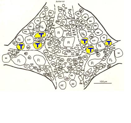
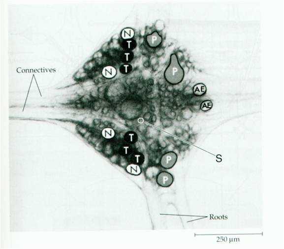
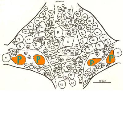
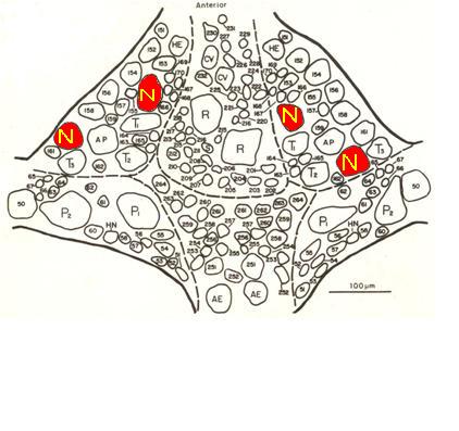
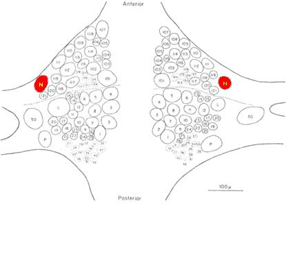
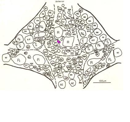
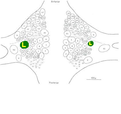
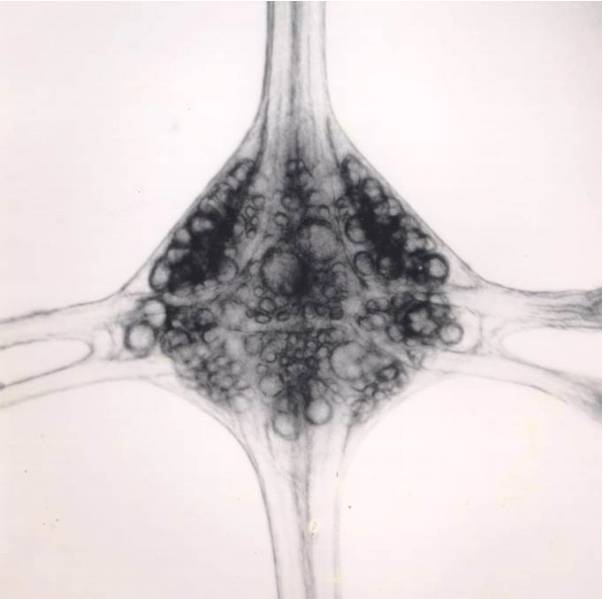
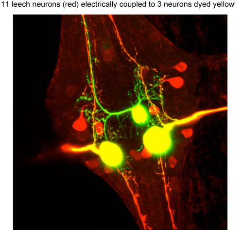
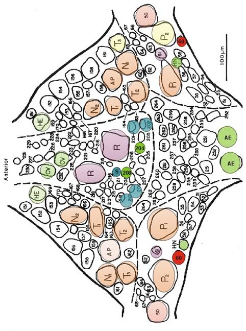
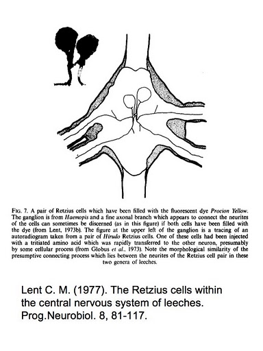
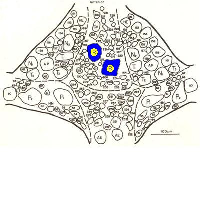
Leech figures
Shortening Neurocircuitry
end Acronyms/Abbreviations
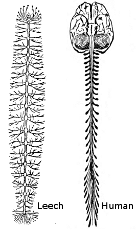

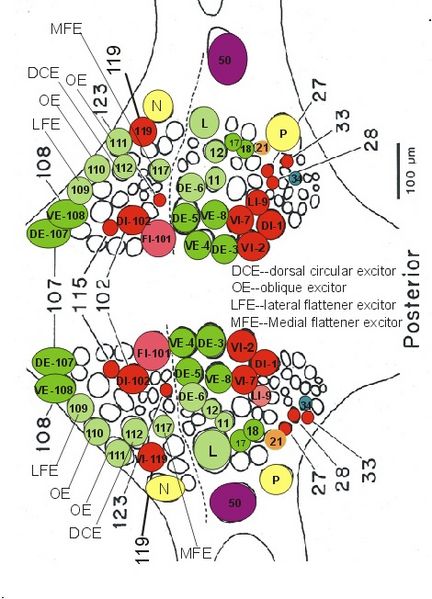
left = anterior Dorsal View (left figure) Ventral View (right figure)
Ventral View (left figure) Dorsal View (right figure)
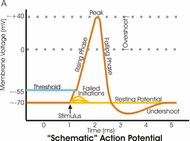
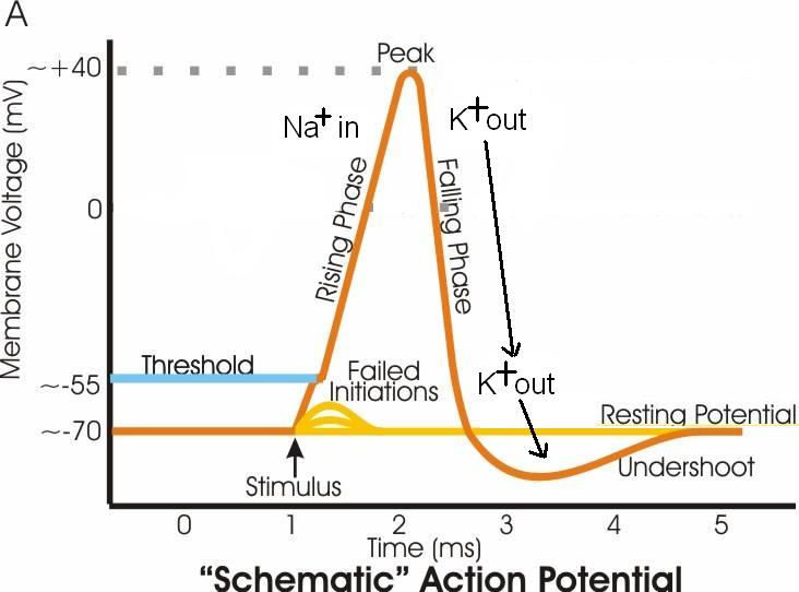
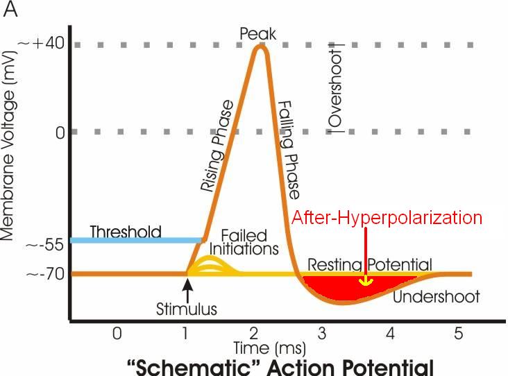
Na+ in first, then K+ out
Afterhypolarization